

The 38 trillion “tenants” in your gut are manipulating your immune system—they are both tenants and business partners.
June 3, 2026
If we could predict exactly how your immune system will respond before a disease manifests?
June 3, 2026Your immune cells have an "aging archive"—and now, we can read it.
Your immune cells have an "aging archive"—and now, we can read it.
—— p16, p21, Telomeres, CD57, SA-β-Gal: The molecular identification code of immune cell senescence
⏱ A One-Minute Read
In the previous articles, we discussed how T cells, B cells, NK cells, and macrophages age over time. But what exactly does "a cell has aged" mean? How do scientists know? Cellular senescence isn't about looking "wrinkled" — it comes with a precise set of molecular markers that can be identified through laboratory testing. These markers are not just academic tools; they are becoming the clinical foundation for "immune age" assessment and the basis for precisely targeting next-generation anti-aging interventions. Understanding these markers gives you a clearer view of: what immune aging is, how to measure it, and why therapeutic interventions should start from these molecules.
Layer 3 | Core Theory: Five Key Markers of Immune Cell Senescence
| Marker | Significance |
|---|---|
| Telomere length shortening | Telomeres shorten with each cell division; leukocyte telomere length is a classic indicator of cumulative division history and degree of aging in immune cells |
| Elevated p16INK4a / p21 expression | Cell cycle inhibitory proteins that drive cells into a permanent non-dividing state (replicative senescence); significantly upregulated in aged immune cells |
| Elevated SA-β-galactosidase (SA-β-Gal) activity | Senescence-associated lysosomal enzyme activity marker; increased activity at pH 6.0 is a classic histochemical marker of senescent cells |
| High CD57 expression (T cells and NK cells) | CD57 is a surface marker of terminal differentiation and replicative senescence; CD57+CD28- T cells are among the most commonly used peripheral blood markers for T cell aging |
| SASP protein profile (plasma or cell culture supernatant) | A combination of pro-inflammatory factors secreted by senescent cells (IL-6, IL-8, TNF-α, MMPs, etc.); plasma SASP protein levels are a liquid biopsy indicator of systemic aging burden |
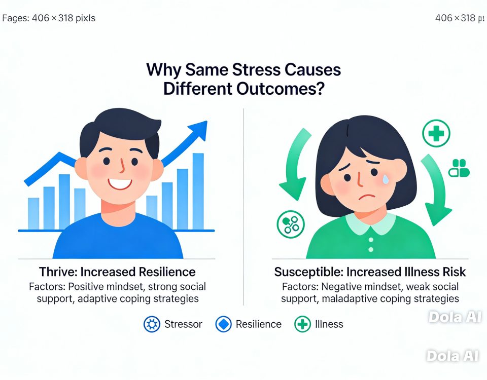
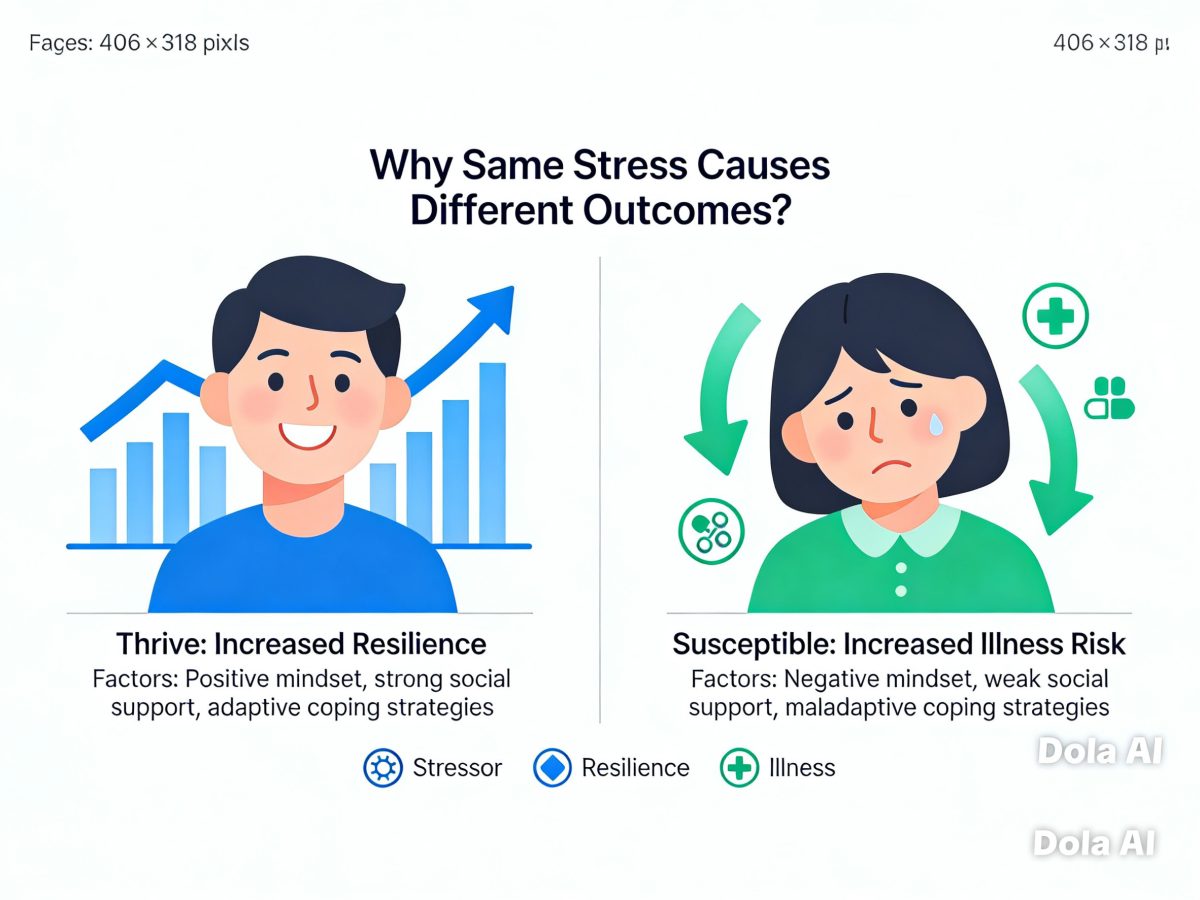
| Core Concept: "Perceived control" is a better predictor of immune impact than objective stress intensity. For two caregivers of a critically ill family member (similar objective stress), the one with sufficient social support and rest opportunities will show significantly different NK cell activity, inflammatory markers, and vaccine efficacy compared to one bearing the burden entirely alone. "Perceived control" is the most critical regulating variable in the stress-immunity dynamic. |
Tier 4 | In-Depth Reading
I. Cellular Senescence: Not dead, but stops working and keeps "shouting"
Before diving into specific markers, let's clarify the concept of "cellular senescence" once more, as it differs somewhat from everyday language about aging.
Cellular senescence is a protective response of cells to certain stressors (telomere shortening, DNA damage, oxidative stress, activation of oncogenes, etc.) — the cell chooses to permanently stop dividing rather than continue proliferating (which might transmit genetic damage) and does not immediately undergo apoptosis (immediate death might cause loss of tissue structure).
In the short term, senescence is beneficial: it prevents cells with genetic damage from replicating indefinitely (cancer prevention) and promotes the secretion of pro-inflammatory signals via SASP to attract immune cells to clear these "problem cells."
However, when the accumulation of senescent cells exceeds the immune system's clearance capacity (which systematically occurs in old age), the continuous SASP output from senescent cells becomes a problem: they become a persistent source of chronic inflammation, damaging surrounding healthy cells and accelerating tissue aging.
Immune cells themselves also undergo senescence — T cells, NK cells, and macrophages all enter a senescent state under long-term antigen stimulation and chronic infection stress. Senescent immune cells, with degraded function but not disappearing, continuously output SASP systemically and are the core cellular source of inflammaging.
Identifying and measuring these senescent immune cells requires a set of specific molecular tools — this is the significance of "senescence markers."
2. Telomere Length: The most intuitive cellular "age counter"
Telomeres are protective repetitive sequences at the ends of chromosomes (repeats of TTAGGG; human telomere length is typically 5–15 kb). With each cell division, DNA replication cannot fully copy the ends of chromosomes, resulting in telomere shortening of approximately 50–100 bp. When telomeres shorten to a critical length (approximately 4–5 kb), the cell senses a "chromosomal end crisis," activates the p53/p21 or p16/Rb signaling pathways, and triggers cellular senescence or apoptosis.
Leukocyte telomere length (LTL) is one of the most widely used biomarkers of immune aging. LTL can be measured from ordinary blood samples using quantitative PCR or Southern blot, and numerous commercial testing services are already available.
Epidemiological data show: the shorter the LTL, the stronger the positive association with all-cause mortality, cardiovascular risk, cancer risk, and neurodegenerative disease risk. Nobel laureate Elizabeth Blackburn's research (and the company she founded, Telomere Diagnostics) has brought telomere length testing into the public eye.
However, interpreting telomere length requires some caveats:
-
LTL reflects the average telomere length of leukocytes as a whole and does not distinguish between different immune cell subsets
-
Telomere length varies greatly between individuals; baseline values are influenced by genetics (approximately 50% of individual differences come from genetics)
-
Rather than looking at a single absolute value, observing the trend over time (whether shortening is accelerating) is more meaningful
-
LTL shortening can be slowed through regular exercise, reducing chronic stress, and smoking cessation — lifestyle effects are real
Telomere length is a physical record of cell division history. Your white blood cell telomeres store information about how many times they have been activated, fought, and proliferated. This "record" can now be read from a tube of blood.
3. p16INK4a: The "Stop Command" Gene for Senescent Cells
p16INK4a (the product of the CDKN2A gene) is a representative cell cycle inhibitor protein. By inhibiting CDK4/6 kinases and preventing the phosphorylation of the Rb protein, it arrests the cell cycle at the G1 phase, leading to permanent cessation of cell division.
In young tissues, the expression of p16 is very low. However, with advancing age, p16 expression increases systematically across almost all tissue types. This upregulation is highly correlated with actual phenotypic aging (e.g., functional decline and increased disease risk).
Research involving genetically engineered mice that target and eliminate senescent cells with high p16 expression has shown that these mice exhibit delayed aging phenotypes (such as muscle atrophy, cataracts, and impaired adipose tissue function) and significantly extended healthspans. Notably, even when this elimination was initiated in old age, beneficial effects were still observed. This experiment established the theoretical foundation for Senolytics (senescent cell-clearing agents) research.
In human immune cells, p16 expression also increases with age, particularly in CD8+ T cells (especially TEMRA cells) and NK cells. Immune cells with high p16 expression lose their proliferative capacity and suffer functional decline, while their secretion of SASP (Senescence-Associated Secretory Phenotype) is enhanced.
It is worth noting that the elevation of p16 is not merely a passive marker of aging; it also serves as a signal that p16 is a "potential target for intervention." One of the mechanisms of action for Senolytics (such as the combination of Dasatinib and Quercetin) is to target the anti-apoptotic pathways of cells with high p16 expression, thereby forcing these senescent cells to re-enter apoptosis and be cleared by the immune system.
4. CD57 and CD28: Flow Cytometry Readouts for T-Cell and NK-Cell Senescence
In both clinical and research settings, flow cytometry is the most widely used tool for immunophenotyping. To assess the senescence status of T cells and NK cells, researchers rely on a standardized combination of surface markers:
For T Cells:
- CD57+CD28- CD8+ T cells: CD57 is a hallmark of terminal differentiation and replicative senescence. CD28 is a co-stimulatory receptor essential for T-cell activation, which is characteristically lost on senescent T cells. This CD57+CD28- phenotype defines a population of "senescent effector T cells" that have essentially lost proliferative capacity but retain cytotoxicity and continuously secrete SASP. The proportion of these cells increases with age and is particularly elevated in individuals who are CMV-positive.
- KLRG1+ (Killer Cell Lectin-like Receptor G1): This is another marker of T-cell terminal differentiation, frequently used in combination with CD57 to refine the senescence profile.
For NK Cells:
- CD57+CD56dim NK cells: CD57 serves as a marker for terminal differentiation in NK cells as well. CD56dim denotes a subset of mature, cytotoxic NK cells; the combined high expression of both markers identifies a population of functionally degraded, senescent NK cells.
- NKG2D Expression Levels: The expression density of the activating receptor NKG2D systematically declines in senescent NK cells, providing a direct readout of their functional status.
These combinations of flow cytometry markers have established a standardized panel for evaluating the "immunosenescence phenotype," which is currently employed in many research medical institutions and precision medicine clinics. institutions and precision medicine clinics.
5. DNA Methylation Clocks: A Systematic Metric for Immunological Age
While telomeres, p16, and CD57 are all single-dimensional markers of senescence, a more systemic tool for assessing "biological age" has recently been emerging into clinical practice: the DNA Methylation Clock (Epigenetic Clock).
DNA methylation is a critical epigenetic mechanism for regulating gene expression; typically, the addition of a methyl group to the CpG sites of a gene suppresses its expression. As we age, the methylation status at specific genomic locations changes in a highly predictable manner. In 2013, Steve Horvath (UCLA) discovered that measuring the methylation status of just 353 CpG sites could predict the "biological age" of a cell, tissue, or individual with remarkable accuracy—a tool he named the "Horvath Clock."
- Subsequent researchers have developed more advanced versions of methylation clocks. Those most relevant to immunosenescence include:
- GrimAge: A biological age metric that incorporates various plasma proteins (including multiple SASP-related proteins). It has been identified as one of the most powerful predictors of all-cause mortality and chronic disease risk.
- PhenoAge: A comprehensive age metric that integrates clinical biochemical markers (such as albumin, white blood cell count, and creatinine).
The most significant application value of methylation clocks lies in their ability to measure the "discrepancy between biological age and chronological age." For instance, some 60-year-olds may have a biological age of only 52 (the opposite of "premature aging"), while others may have a biological age of 70 (accelerated aging). This deviation is one of the most potent indicators for predicting future health risks and acts as a highly sensitive readout for the efficacy of interventions; lifestyle modifications (such as exercise, dietary changes, and improved sleep) can measurably slow the progression of these methylation clocks.
DNA methylation clocks transform the question, "How old is your immune system?" from a vague notion into a precise, quantifiable number. This is more than just an academic tool—it is becoming the new language for personalized immune health management in precision medicine.
6. Practical Applications of Senescence Markers: What Can Be Tested Now?
For individuals seeking to proactively understand their immune aging status, the following tests are currently available in practice:
- Leukocyte Telomere Length (LTL): Offered by numerous commercial laboratories and some functional medicine clinics. Based on blood samples, these reports compare an individual's telomere length to the percentile of the general population within their age group.
- Inflammatory Marker Panels (hs-CRP, IL-6, TNF-α): These reflect the system-wide inflammatory burden associated with SASP. These are often included in comprehensive health screening packages or can be requested as individual tests.
- Flow Cytometric Analysis of Immune Cell Subsets: At medical institutions with specialized immunology departments, one can request flow cytometry to analyze T-cell subsets (including the CD57+CD28- population) and NK-cell activity (NKCA).
- DNA Methylation Biological Age (Epigenetic Clocks): Some functional medicine clinics and longevity-focused medical institutions have begun offering methylation clock tests (such as the TrueDiagnostic TruAge panel). While still relatively expensive, the cost is gradually decreasing.
Currently, these tests are primarily utilized by individuals with a high degree of proactive health management awareness rather than being included in standard annual physicals. However, as testing costs decline and clinical evidence continues to accumulate, it is only a matter of time before they are incorporated into routine annual check-up packages.
Knowing your "immunological age" and using it to guide targeted lifestyle optimization and medical interventions is the fundamental logic of precision immune health management—a far more reliable approach than subjective assessments based on "feeling pretty good."
Figure: Five Key Marker Systems for Immune Cell Senescence
| Marker | Methodology |
|---|---|
| Telomere Length (LTL) | qPCR / Southern blot (Blood) |
| p16INK4a Expression | Immunohistochemistry / qPCR (Tissue/Blood) |
| CD57+CD28- T Cells | Flow Cytometry (Blood) |
| NK Cell Activity (NKCA) | in vitro Cytotoxicity Assay (Blood) |
| DNA Methylation Clocks (e.g., GrimAge) | Methylation Array/Sequencing (Blood) |
Key Takeaways
-
Cellular senescence is not death; rather, it is a state of permanent growth arrest characterized by the secretion of pro-inflammatory factors known as SASP. While this mechanism serves as a safeguard against cancer, its accumulation acts as a primary driver of chronic inflammation.
-
Leukocyte Telomere Length (LTL) is the most widely utilized biomarker for immune aging. Accessible via standard blood tests, it reflects the cumulative division history of leukocytes and is highly correlated with the risk of chronic diseases.
-
p16INK4a functions as the molecular "switch" that drives cells into permanent senescence. The targeted clearance of these cells (Senolytics) represents one of the most promising avenues for anti-aging interventions.
-
The CD57+CD28- T-cell ratio and NK Cell Activity (NKCA) serve as the most direct, measurable flow cytometry readouts for immune functional decline and are currently available at specialized immunology centers.
-
DNA Methylation Clocks (particularly GrimAge) quantify "biological age" into precise, measurable data, serving as one of the most powerful tools for predicting chronic disease risk and evaluating the efficacy of health interventions.
-
Determining your "immunological age" is the starting point for precision immune health management. A multi-dimensional assessment combining various biomarkers is significantly more reliable and comprehensive than relying on any single indicator.


